Three ulnar-innervated hand muscles are commonly used for train-of-four monitoring. Emerging evidence shows they don’t all behave the same.
With the release of the 2023 Practice Guidelines for Monitoring and Antagonism of Neuromuscular Blockade from the American Society of Anesthesiologists, anesthesia departments are now expected to routinely use quantitative neuromuscular monitoring whenever non-depolarizing neuromuscular blocking agents are administered.
The goal of these guidelines is clear: Confirm adequate recovery (TOF ratio ≥ 0.9) prior to extubation to reduce postoperative respiratory complications.
Importantly, the guidelines were delayed for years—not due to lack of evidence—but due to concerns that older quantitative monitors were too difficult, unreliable, or disruptive to routine workflow. As summarized in the 2018 international consensus statement:
“A major limitation to widespread adoption of objective neuromuscular monitors is not their cost, but their ease of use… New monitors that are reliable, easy to set up, and produce repeatable responses are needed.”1

Figure 1: A mechanomyograph as shown in a 1976 Anesthesiology article5.
The 2023 guidelines acknowledge that this barrier is now changing with the introduction of substantially improved technology.2
Key takeaway:
Before comparing features or prices, ensure the device can reliably support guideline-level decision-making in real clinical conditions.
Step 2: Understand What All Quantitative Monitors Are Trying to Measure
All quantitative neuromuscular monitors share the same physiological goal: Measure the muscle’s response to peripheral nerve stimulation and express recovery as a Train-of-Four (TOF) ratio. As demonstrated by Eikermann, even modest levels of residual neuromuscular blockade (TOF ratios as high as 80%) may cause significant airway narrowing3.

Figure 2: Minimum retroglossal upper airway diameter during forced inspiration (A) before neuromuscular blockade (baseline), at a steady-state TOF ratio of (B) 0.5 and (C) 0.8. Images from the volunteer show that a partial paralysis evokes an impairment of upper airway diameter increase during forced inspiration. *p < 0.05 versus baseline.3
What differs is how that response is detected.
Historically and currently, the following technologies have been used:
Why this matters:
MMG is the reference gold standard used to define TOF ≥ 0.9 as adequate recovery. Any monitor used clinically should produce results that are equivalent to MMG4.
MMG established the science of neuromuscular recovery but required rigid arm fixation, preload weights, and specialized setup—making it unsuitable for routine OR use.
To overcome MMG’s impracticality, many first-generation clinical monitors adopted acceleromyography (AMG). While easier to deploy, AMG introduced two major problems5:
As summarized in the referenced literature:
Key takeaway:
A monitor that requires manual correction or ideal limb positioning undermines the very safety goal it is meant to support.
Advances in electronics and signal processing have enabled second-generation EMG-based monitors, which overcome many historical limitations of AMG.
Advantages of modern Electromyography:
However, EMG introduces a new challenge: electrical noise.
The EMG signal is small and exists in an electrically noisy OR environment. Poor signal handling can lead to:
Experts have therefore emphasized:
“The performance of commercially available twitch monitors should be compared to mechanomyography, using mechanomyography as the reference gold standard.”5
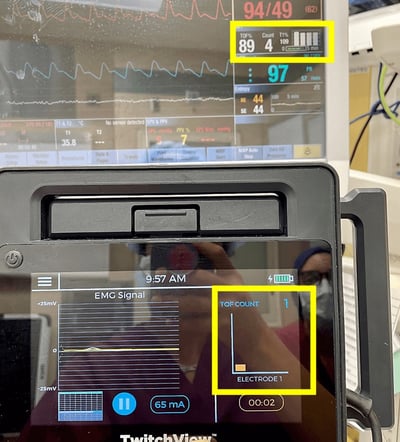
%20monitoring%20devices..png?width=3046&height=1922&name=Simultaneous%20measurements%20from%20two%20electromyography%20(EMG)%20monitoring%20devices..png)
Figure 3: Simultaneous measurements from two electromyography (EMG) monitoring devices. TwitchView portrays expected linear recovery while EMG Monitor 2 underestimates recovery. EMG monitor 2 fails to measure post-tetanic count (PTC) and undercounts twitches.7
Key takeaway:
Choosing “EMG” is not enough. The system must be clinically validated against MMG across all depths of blockade.
Not all EMG monitors are validated under conditions that reflect routine clinical practice. Some devices demonstrate performance in controlled laboratory or research environments, where patient positioning is ideal, electrical interference is minimal, and monitoring is managed by trained investigators rather than busy clinical teams.
While such testing may establish technical feasibility, it does not necessarily predict reliability in the operating room, where electrocautery, warming devices, patient movement, variable electrode placement, and time pressure are routine.
Clinically meaningful validation requires demonstration that a monitor provides stable, interpretable data across a range of patients and surgical conditions, when used by typical anesthesia providers, and incorporated into standard workflows. Without evidence of performance in real-world OR settings, technical specifications alone are insufficient to support adoption as a routine clinical tool.
What counts as clinical validation?
When evaluating a quantitative neuromuscular monitor, look for evidence that it has been:
If these criteria are not met, technical specifications alone are unlikely to predict real-world reliability.
Finally, technology only improves outcomes if it is used consistently.
Despite access to quantitative monitors, many clinicians skip using them, relying on sugammadex as a safety net instead.
However, a large database study showed that while sugammadex reduced residual neuromuscular blockade compared with neostigmine—complete elimination of respiratory complications required quantitative neuromuscular monitoring.8
Independent studies of universal quantitative monitoring show why this matters. In one academic medical center analysis, 60% of patients judged ready for extubation using qualitative assessment still had a TOF ratio < 0.9—placing them at risk for postoperative pulmonary complications.9
Even modest reductions in postoperative pulmonary complications were projected to more than offset the cost of universal quantitative monitoring, making accuracy not only a clinical issue—but an operational one.
When evaluating monitors, ask:
Ease of use was explicitly cited as the primary barrier to adoption—not cost.
When choosing a quantitative neuromuscular monitor, prioritize:
The 2023 ASA guidelines marked a turning point. Quantitative neuromuscular monitoring is recommended as the standard of care—but not all monitors are equally capable of delivering the safety the guidelines demand.
Choosing the right technology isn’t just about buying a monitor—it’s about selecting one that provides all the data that is needed and can be easily accessed and used.
When evaluating a new monitor, ask yourself “Can I trust what the monitor is telling me at the moment it matters most?”
Learn why the TwitchView® Train of Four Monitor sets the gold standard for EMG based monitors.8 9
TwitchView® has been clinically validated in several independent, third-party hospital settings, where performance is assessed under routine clinical conditions. These validations extended beyond technical noise cancellation for accurate EMG signal detection to clinically relevant measures, including customized management of neuromuscular blockade through trended data, guidance of reversal agent selection, and analysis of postoperative pulmonary outcomes and cost of care.9 10 11